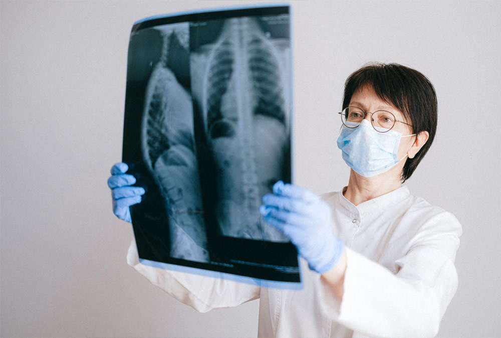
Radiology is the fastest developing branch of medicine, and has two divisions- Radiodiagnosis and Radiotherapy. Radiology service involve the Radiologists as well as Radiographers;so as well trained Radiographers should be available for efficient radiology work. The criteria for selection and training should be formulated with this in mind. Diagnostic radiology performed with Roentgen rays.
| Eligibility | The Minimum Qualification Is S S L C but Preference Will Be Given To +2 With Science Subjects. Both Men And Women Are Admitted. DIPLOMA Will be Issued Only to Successful Candidates |
| Duration | 2 Years |
| Diagnostic Radiology(X-Ray) & Other options | 1000 mA & 300 mA X-Ray Machine -1 C-Arm With Image Intensifier-1, E C G -2 C T, Cath Lab-(Optional) |
| Maximum Student Strength | 20 Numbers |
| Medical Institute | Medical Institute affiliated to KPPMA (Kerala State) Navajyothy Medical Institute with maximum student of 20 strength. |
| Class Rooms (25x10sq ft) | 5 |
| Multimedia | Projector with laser Pointer |
| Library | Minimum 100sq ft With Computers & Internet facility Minimum of 100 books |
| Teaching facilities | Part time Lecturers ,cardiologist, Radiologists (Conventional and Interventional) Ortho specialist, ENT specialist, Pediatrics ,Radiology Technician (Tutor) etc. |
| Final Year Examination | Theory Examination + Practical +Record +Viva voce |
| Academic Fee | Rs.1.5 Lakhs |
| Inspection authority | Director of Radiation safety (D R S)Kerala, issued by the institute. |
| Main Subjects | |
| 1st Year (Semester I & II ) | |
| 1 | Anatomy and Physiology |
| 2 | Basic Physics. |
| 3 | Electrical and Radiation Physics. |
| 4 | Positioning in Radiography and Topographic Anatomy |
| 5 | Patient care during radiographic examination |
| 6 | Principles of Radiographic Exposure and Medical Emergencies |
| II nd Year (Semester III & IV) | |
| 7 | Radiation protection |
| 8 | Special X-Ray investigations |
| 9 | Dark room chemistry and techniques. |
| 10 | Pediatric Radiography. |
| 11 | Medical-Surgical Diseases |
| 12 | Ethics and Supervised film critique sessions |
NOTE: Communication medium: English
All the subjects with practical included in the final year examination and issue the certificate to the successful candidates.
| Patron | Dr Jones P Mathew MBBS |
| Director | Asokan A P Bsc DRT (Radiographer) |
| Dept. of Radiology | Dr C P Mathew MBBS, MS, DMRD, DMRT (Rtd. Prof. of Radiology Government Medical College Kottayam) |
The Hostellers Administration And Discipline at The Hostels Are Governed by Hostel Rules. The Hostellers Are Responsible to Maintain all Furniture, Fan, Bulbs, Electrical Fittings, Sanitary Fittings etc…. Their Respective Room and Hostel.
The Institute is Located at The Centre of Vaikom, Near Boat Jetty By Walk, 5 Minutes from K.S.R.T.C.Bus Stand.Vaikom Is located 33 Kilometers Away From Ernakulum Railway Station. From Ernakulum K.S.R.T.C. Bus Stand There Are Buses to Vaikom And Via Vaikom.
| Anatomy and Physiology | 300 Hours |
| Basic Physics. | 150 Hours |
| Electrical and Radiation Physics | 300 Hours |
| Positioning in Radiography, Topographic Anatomy. | 350 Hours |
| Patient care during radiographic examination. | 250 Hours |
| Exposure Principles of Radiographic | 50 Hours |
Typical cell Tissues structure of skeletal system. Bones and ossifications, Joints of upper limb, lower limb, shoulder girdle, pelvic girdle, vertebral column , thorax, skull and Locomotor system, skeletal system, Circulatory system, Respiratory system, Lymphatic system, Reticulo- endothelial system, Digestive system, Genito-urinary system, Muscular system, Endocrine system, Nervous system, Cranial Nervesand CSF, Special Sense Nerves organs: Eye, Ear, Tongue, Surface markings and Cell function, Function of tissues. Function of bones and joints. Physiology of circulation, Physiology of respiratory system. Function of Lymphatic system. Function of Pituitary, thyroid, parathyroid , adrenal, thymus, pancreas and gonads. Function of brain and the nerves. Function of the eye and the ear. Functions of Reproduction system and pregnancy rule, method of Family Planning.
Measurements and Units, heat,Energy and Mechanics, Electricity and Magnetism, laws of magnetic force, electrostastics, conductors and insulators ,capacity and condenser DC, AC.. Basic electronics. Two phase and three phase,Transformer s (Step up and step down) Rectification (Two wave and three wave) ampere, coulomb, electromagnetic induction ,line Voltage, power, RMS values, Diodes ,ammeters and volt meters, Circuits, Ohms Law and Faraday’s Law,solenoids
Electro-magnetic radiation ,Electro Magnetic Spectrum, Production of X-rays, Thermionic emission, ionization by Xrays, roentgen, target material ,Coolidge tube, Properties of X-rays,cooling of Xray tube, Interaction of Radiation with matter, induction experiments, Radioactivity, Measurement of Radiation, ionization measurements, radiation units ,R B E, LET,Biological effects of radiation, Radiation protection, Code of practice, Protective materials and methods, Personnel monitoring.(Film badge and T L D) Cassettes ,Films and Grid (Stationary ,rotating, parallel ,crossed, focused pseudo focused.)
It Includes Supervised Theoretical and Practical Study of Positioning of patients For different Radiological Projections and special views. Different radiological baselines, different Surface anatomy including vertebral levels and boney projections. Erect supine and oblique positions, care and management of X ray tubes, positioning of causality patients, care of pre and post surgical patients, positioning for portable machines, infants and children, different tube angulations. Identification of the side of patients
Radiation protection ,shielding, Hospital staffing and organization. Records relating to patients and departmental statistics, Professional attitudes. Communication. Medico legal aspects. Appointment Units. Patient psychology. Counselling, Management of Wheel chair and stretcher patients. Nursing care. First aid. Care of unconscious patients. Preparation of patients for X-ray examinations. infection control. Sterilization ,Emergency, Drugs in the X-ray department, Contrast agents. Hypersensitivity and reaction to contrast agents, Medical Terminology, Management of medical hazards patients. Hygiene and sterility, dressing code of male and female patients. Patients with supporting medium, fracture and dislocations
Radiation protection, time of exposure, milli ampearage, transformers, tube current ,anode and cathode , shielding, vacuum tube ,thermionic emission, properties of X-rays filtration, HVL. coolidge tube, cooling of X ray tube, collimation, heating, protective barriers, secondary radiation, primary beam, grids, insulation, room lay out. time management, identification of different errors and their rectifications, exposure control with respect to the patients condition
| Second year Semester 1500 Hours and Practical 900 Hours | |
| Subjects & Number of hours | |
| Radiation protection | 150 Hours |
| Special X-Ray investigations. | 400 Hours |
| Dark room chemistry and Techniques. | 400 Hours |
| Pediatric Radiography. | 250 Hours |
| Medical-Surgical Diseases. | 100 Hours |
| Ethics and Supervised Film critique sessions. | 100 Hours |
Principles of radiation protection, shielding and collimation, different gonad shields, lead aprons, secondary protection barriers ,time, principles of radiation attenuation, filters. aluminum filters, Half value layer, Radiation monitoring devices. distance management, gonads protection, ten day rule, secondary lead barriers, shielding of radiographic and dark rooms, measurement of radiation exposure ,care of younger patients, protection of the public, alarm and monitoring devices, protection symbols, cubicle, protection of unexposed films and cassettes, warning signs and safety boards
Special radiological procedures ,intravenous pylogram, micturating cysto urethrogram,carotid angiogram ,cerebral angiogram,coronary angiogram,Angioplasty,Discography.oral cholecystogram,cholangiography,Myelography,Sialography,Barium swallow,Barium meal follow through,Barium enema,Double contrast study,hysterosalphingography, percutaneous transhepatic cholangiography, Dacryo-cystography Digital substaction angiography, bronchography, Lymphangiography ,Arthrography, pelvimetry, cephalometry,mammography, fluoroscopy,cineradiography,contrast agents (ionic and non ionic), emergency medicines, contrast reactions.
Chemicals for developer and fixer, tanks,temperature and humidity control, pH values,density, hardening,developing and fixing of exposed films,chemical processing, changing of chemicals,washing, hangers,dryers, intensifying screens and their interactions with Xrays, cassettes,safelights and room lights,dark room adaptations,red goggles.Lay out of Dark room, Wet and Dry areas, care and management of unexposed films, cleaning of hangers and screens, statistics of films , introduction of automatic processing
Radiation protection of infants, invertogram, care of infants ,oral and local anesthesia, first aid ,topographic anatomy, managements of children with fracture and dislocations, special skull projections, chest Xray management ,pre and post surgical patients, care of neonates, portable radiography, different erect views, shields, incubator, variations in the exposure factors, size`of films, counseling
Common pathological terms, shock,inflammation,infections,neoplasms,disorders,tumours (benign and malignant) infarcts,ulcers and necrosis, gout,rheumatism, embolism, haemophilia, HIV paralysis,neuralgia,sarcoma and carcinoma , tracheoestomy and colostomy patients, patients with emergency tubes, pre and post surgical patients, patients with metallic supports, pacemaker,vertebral dislocations and fractures, amputation,
Medical ethics ,radiological ethics, guidelines , daily management of critical patients,storage of films,exposed and un exposed films ,statistics of different films, loading and unloading of films.cleaning,
During two year the students are placed in x-ray departments on a alternate basis to gain practical experience in relevant areas under supervision of Tutor radiographers and the senior radiographers.
They have to maintain a record of their work in the ‘Student Record of Practical work” provided by the school.
Written test conducted at end of First and second semesters, And third and fourth semesters.
Practical exam and viva examination at end of First and second semesters, And third and fourth semesters
80% attendance is a must to appear for the final examinations. Periodic evaluation should be done every semester both in theory and practical. 15% of final exam should be from intern assessment and rest 85% from theory and practical.
| Sessions | No. of questions | Marks per question | Total Marks |
| Essays | 3 | 10 | 30 |
| Short Notes | 8 | 5 | 40 |
| One ward Answer | 15 | 2 | 30 |
| Total | 100 | ||
| Part | Theory | Practicals & Viva |
| PAPER-I | Anatomy and Physiology | Identification of Bones and Structures |
| PAPER-II | Basic Physics, | Identification of Electrical Equipments |
| PAPER-III | Electrical and Radiation Physics | Equipments related to X-Ray Exposure |
| PAPER-IV | Positioning in Radiography and Topography Anatomy | Positioning for different Radiological Projections |
| PAPER-V | Patient care during Radiographic examination | Radiation Protection and Management |
| PAPER-VI | Principles of Radiographic Exposure | Factors ,KVP , mAs and Protective Barriers |
| PAPER-VII | Radiation Protection | Shielding ,collimation and patient care |
| PAPER-VIII | Special X-Ray investigations | Contrast agents, special radiological procedures |
| PAPER-IX | Dark room chemistry and Techniques | Developing, Fixing ,Washing and dark Room Adaptations |
| PAPER-X | Pediatric Radiography | Management Of Infants, Different Radiographic Projections Of Children |
| PAPER-XI | Medical-Surgical Diseases | Management Of Pre and Post Surgical Patients |
| PAPER-XII | Ethics and Supervised film critique sessions | Ethics of Radiology |
| Theory | 100 |
| Practical | 150 |
| Viva & Voice | 50 |
| Intern assessment | 50 |